Provided at OPENSCREEN-GR by
- NCSR “Demokritos” Athens
- Democritus University of Thrace

Services
Optical Microscopy
Optical Microscopy - NCSR “Demokritos” Athens
The Optical Microscopy Unit of the Institute of Biosciences and Applications (IBA) of NCSR “Demokritos” offers state-of-the-art equipment, which serves imaging needs of the research laboratories of the Institute as well as hospitals and universities
Equipment:
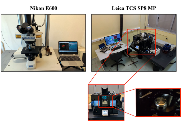
Leica TCS SP8 MP
A multi-photon confocal microscope with a fully automated motor bank. The system is accompanied by a incubator chamber for the strict control of all environmental conditions (humidity, temperature, CO2, O2, N2). Multi-image / confocal microscopy system (Leica SP8 in inverted DMi8, equipped with PMT and two hybrid detectors, three AOTF-regulated laser sources, a Mai Tai eHP Deep See laser and climate chamber)
BioRad MRC 1024
Confocal microscope equipped with a Nikon E600 upright optical microscope (3 color detection).
Applications:
This unit covers a wide range of optical microscopy applications, such as:
- Multi-channel Fluorescence Microscopy, covering the ultraviolet, visible spectrum and infrared
- Multicolor 3D Imaging
- Live cell imaging Two-photon confocal microscopy
- Second Harmonic Generation imaging protocols
- Förster [Förster / Fluorescence Resonance Energy Transfer (FRET)] protocols for monitoring molecular interactions in living and permanent cells
- Photofluorescence Fluorescence Recovery Protocols (FRAP) Co-location analysis in live and fixed cells
- Calcium ion imaging (Calcium imaging)
- Spectral Unmixing Differential Contrast Contrast Microscopy (DIC) (known as Nomarski Microscopy)
- Image processing and analysis (with specialized software such as ImageJ / Fiji and Imaris (Bitplane)
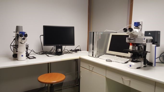
Microscopy System - Democritus University of Thrace
Equipment:
The unit has a fluorescence microscope (ZEISS Scope.A1) equipped with a camera (Axiocam ICm1) and connected to a computer with the ZEN 2 image analysis program (ZEISS). The unit also has a fluorescence microscope (ZEISS Scope.A1) equipped with a camera (Axiocam ICm1) and connected to a computer with the ZEN 2 image analysis program (ZEISS).
Applications
- Multi-channel fluorescence microscopy
- Imaging / videotaping of live cells at consecutive intervals
- In vitro analysis of cell migration
- Analysis of oxidative damage (comet assay)
- Phase contrast microscopy
- Differential Contrast Contrast Microscopy (DIC-Nomarski)
- Digital image processing, analysis and quantification